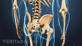
The hip’s unique anatomy enables it to be both extremely strong and amazingly flexible, so it can bear weight and allow for a wide range of movement.

The hip is located where the head of the femur, or thighbone, fits into a rounded socket of the pelvis. This ball-and-socket construction allows for three distinct types of flexibility:
- Hip flexion and extension - moving the leg back and forth;
- Hip abduction and adduction - moving the leg out to the side (abduction) and inward toward the other leg (adduction); and
- Rotation - pointing toes inward (internal rotation) or outward (external rotation) and then moving the straightened leg in the direction of the toes.
Also known as the acetabulofemoral joint, the hip joint is comprised of these basic components:
- Hip bones, including the femur and pelvic bones;
- Hip articular cartilage that decreases friction between the bones and allows for a smooth gliding motion;
- Hip muscles that both support the joint and enable movement;
- Hip ligaments and tendons, tough, fibrous tissues that bind bones to bones and muscles to bones; and
- Synovial membrane and fluid, which encapsulates the hip joint and lubricates it, respectively.
Hip problems occur when any one of these components starts to degenerate or is in some way compromised or irritated.
Structure of the Hip Joint
The hip joint is constructed and functions as follows:
Hip bones
- The hip is located where the top of the femur bone, or thighbone, fits into the pelvis.
- The femur bone is the longest bone in the body, extending from the knee to the hip. At the top of the femur bone is the greater trochanter, that boney knob that people often refer to as their hip, and the bulbous "femoral head" which makes contact with the pelvis and forms the ball of the ball and socket joint. It is covered with articular cartilage, which serves to decrease friction.
- The femoral head fits into the acetabulum, the round socket of the pelvic bone.
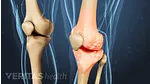
Hip articular cartilage
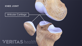
- Both the femoral head and the acetabulum are lined with articular cartilage.
- Articular cartilage is an extremely slippery, strong, flexible material that covers a bone at the site of the joint.
- When the femoral head rotates in the acetabulum, the articular cartilage allows the two surfaces to glide against each other.
- Articular cartilage also acts as a shock absorber, cushioning bones against impacting each other (e.g. while jumping).
- When articular cartilage is damaged or thinned, its ability to protect against friction and impact are impeded.
Read more about What Is Cartilage?
- A strong piece of cartilage, called the labrum, rings the outer edge of the acetabulum. The labrum deepens the socket joint, making the joint more stable, but its elasticity allows for flexibility.
- Over time the smooth articular cartilage that makes up the surfaces of the hip joint may be damaged or blemished by normal wear and tear or from trauma. This process is referred to as arthritis.
Hip ligaments and tendons
- The hip has several ligaments connecting the femur to the pelvis and tendons connecting the bones to many surrounding muscles. Tightness of these ligaments and tendons can cause hip instability and pain.
- Strong and flexible ligaments and tendons provide structure to the hip, reducing strain on the joint, so most hip osteoarthritis therapies will including stretching and strengthening of these structures.
Synovial fluid
- A synovial membrane encapsulates the entire hip joint. This membrane produces synovial fluid, a viscous substance that lubricates and circulates nutrients to the joint.
- When the hip is at rest, the synovial fluid is stored in the cartilage, much like water in a sponge.
- When the hip rotates or bears weight the synovial fluid is squeezed out. Therefore, joint use is necessary to keep the hip joint lubricated and healthy.
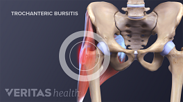
- Small sacs of synovial fluid, called bursae (singular is "bursa") surround all major joints, including the hip joint. Bursae help protect the muscles, bones and tendons from friction as the hip moves. When the bursae are inflamed, the condition is called hip bursitis.
Hip muscles
The hip joint is surrounded by several muscles, including:
- Gluteal muscles, located on the back of the hip (buttocks);
- The adductor muscle on the inner thigh;
- The iliopsoas muscle, which extends from the lower back to upper femur;
- Quadriceps, a group of four muscles that comprise the front of the thigh; and
- Hamstrings, a group of muscles that comprise the back of the thigh and extend to just below the knee.
Together, these muscles support the hip joint, so exercises to relieve hip osteoarthritis symptoms will focus on these muscles as well as muscles of the core.
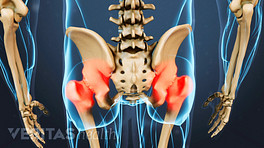
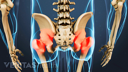
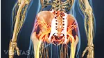
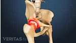
When the hip joint becomes inflamed and painful, the pain may be felt in the groin but may also be referred into the back, buttocks and down the front or back of the leg.
Hip Anatomy on X-Rays and Other Imaging Studies
It is important to note that the degree of anatomical problems (such as the degree of hip joint deterioration) that can be seen on a hip x-ray does not always correlate with the level of pain.
For example, a patient could have hip x-rays that show moderate right hip arthritis and mild arthritis on the left, but he or she may only have pain on the side showing mild hip arthritis.
This counterintuitive result is due to the fact that the pain felt from arthritis is usually due to inflammation in the joint and not the arthritis itself. This concept applies to all the joints in the body and is important to understand because treating the inflammation will often treat the pain and dysfunction and can often help someone put off surgery or avoid it all together.