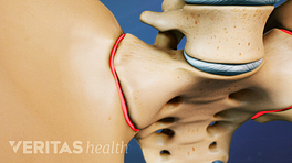
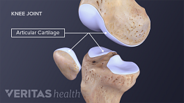
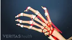
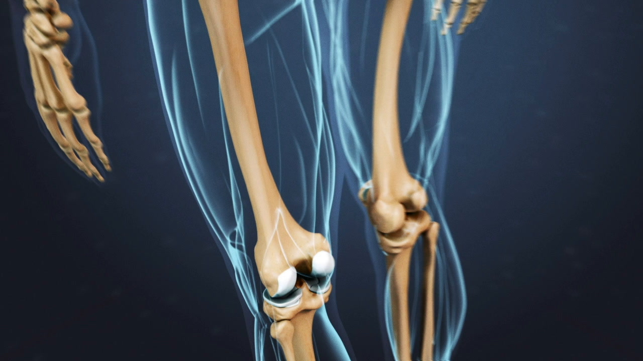
When a healthy joint moves, its bones glide against one another with little or no friction. This ease of motion exists because the boney surfaces are buffered by:
- A layer of slick articular cartilage that can measure anywhere from less than 1 mm to more than 6 mm thick 1 Frisbie DD, Cross MW, McIlwraith CW. A comparative study of articular cartilage thickness in the stifle of animal species used in human pre-clinical studies compared to articular cartilage thickness in the human knee. Vet Comp Orthop Traumatol. 2006;19(3):142-6. PubMed PMID: 16971996. , 2 Pollock J, O'Toole RV, Nowicki SD, Eglseder WA. Articular cartilage thickness at the distal radius: a cadaveric study. J Hand Surg Am. 2013 Aug;38(8):1477-81. doi: 10.1016/j.jhsa.2013.04.037. Epub 2013 Jun 28. PubMed PMID: 23810572. , 3 Cohen ZA, McCarthy DM, Kwak SD, Legrand P, Fogarasi F, Ciaccio EJ, Ateshian GA. Knee cartilage topography, thickness, and contact areas from MRI: in-vitro calibration and in-vivo measurements.
- 0.15 to 4 mL of slippery synovial fluid 4 Mundt L, Shanahan K MS MT(ASCP), Graf’s Textbook of Routine Urinalysis and Body Fluids (p.255), Philadelphia, Pa: Lippincott Williams & Wilkins; 2011 , 5 Osteoarthritis Cartilage. 1999 Jan;7(1):95-109. PubMed PMID: 10367018.
In This Article:
- What Is a Synovial Joint?
- How Do Synovial Joints Work?
Synovial fluid's primary job is to provide cushion and lubrication for joints. A joint's synovial membrane produces substances called albumin and hyaluronic acid that give the synovial fluid its viscosity and slickness. In addition, synovial fluid delivers nutrients to the cartilage and removes waste from the cartilage.
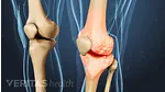
Healthy Joints Need to Move
When a joint is at rest, cartilage absorbs some of the synovial fluid. When the joint is in use the synovial fluid is squeezed out of the cartilage, much like how water is wrung from a sponge. Consequently, joint use is essential to circulate the synovial fluid throughout the joint.
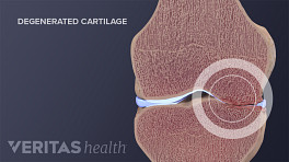
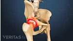
Arthritis and Injuries in Synovial Joints
When the cartilage is damaged as in osteoarthritis, the body responds by increasing the production of synovial fluid (sometimes 3 times as much) in order to compensate for the diseased joint. The excess fluid can cause the joint to become distended, causing further pain.
See Osteoarthritis Symptoms and Signs
In addition to producing synovial fluid, the synovial membrane acts as a semi-permeable barrier for the joint, allowing certain types of cells to flow in and out of it. For example, a traumatic knee injury, such as an anterior cruciate ligament (ACL) tear or a bone break at the knee joint, can cause the synovial joint capsule to fill with blood within minutes or hours of the injury. As a joint recovers, the blood will dissipate and the joint fluid will return to normal.
Fibrous Joints and Cartilaginous Joints
In addition to synovial joints, the adult body contains two other types of joints.
- Fibrous joints occur where two bones are joined by fibers. For example, the seams of the human skull are fibrous joints that show where skull bones grow together during infancy.
- Cartilaginous joints occur where two bones are joined by cartilage, such as where the first rib meets the sternum.
Because of their structural simplicity and because they allow for little or no movement, these two types of joints are generally less prone to injury and problems such as arthritis.
- 1 Frisbie DD, Cross MW, McIlwraith CW. A comparative study of articular cartilage thickness in the stifle of animal species used in human pre-clinical studies compared to articular cartilage thickness in the human knee. Vet Comp Orthop Traumatol. 2006;19(3):142-6. PubMed PMID: 16971996.
- 2 Pollock J, O'Toole RV, Nowicki SD, Eglseder WA. Articular cartilage thickness at the distal radius: a cadaveric study. J Hand Surg Am. 2013 Aug;38(8):1477-81. doi: 10.1016/j.jhsa.2013.04.037. Epub 2013 Jun 28. PubMed PMID: 23810572.
- 3 Cohen ZA, McCarthy DM, Kwak SD, Legrand P, Fogarasi F, Ciaccio EJ, Ateshian GA. Knee cartilage topography, thickness, and contact areas from MRI: in-vitro calibration and in-vivo measurements.
- 4 Mundt L, Shanahan K MS MT(ASCP), Graf’s Textbook of Routine Urinalysis and Body Fluids (p.255), Philadelphia, Pa: Lippincott Williams & Wilkins; 2011
- 5 Osteoarthritis Cartilage. 1999 Jan;7(1):95-109. PubMed PMID: 10367018.