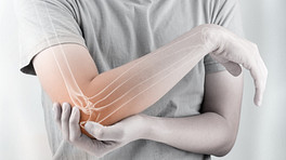
The sooner rheumatoid arthritis is accurately diagnosed, the sooner treatment can be started to limit its effects. Diagnosing rheumatoid arthritis can be challenging, though. A person may have multiple appointments with a primary care physician and specialists before a diagnosis is made.

RA signs and symptoms vary so much that the disease may be hard to recognize. For example, one person may develop puffy, stiff wrist and finger joints over many months, while another person may develop fatigue, fever, and a severely inflamed knee almost overnight. In addition, there is no single lab test that can positively diagnose RA. To help with this challenge, most physicians follow a set of clear guidelines and a point system.
In This Article:
Physician Expertise
Because diagnosing rheumatoid arthritis can be challenging, experts recommend making an appointment with a rheumatologist or a physician who has a lot of experience with inflammatory arthritis. He or she will be familiar with the signs and symptoms of RA and similar diseases, such as psoriatic arthritis, lupus, and reactive arthritis.
See Rheumatologist for Arthritis Treatment
Undifferentiated Arthritis
If a person does not meet the diagnostic criteria for RA or another disease, they may be diagnosed with what doctors call undifferentiated arthritis. Of those diagnosed with undifferentiated arthritis 1 Riaz, S and Kontzias, A. Rheumatoid Arthritis. In: Efthimiou P, ed. Absolute Rheumatology Review. Springer Nature Switzerland AG; 2020; chap 15. Accessed September 15, 2020. https://doi.org/10.1007/978-3-030-23022-7_15 :
- 33% will later be diagnosed with RA (signs and symptoms get worse)
- 33% will later be diagnosed with another disease, such as reactive arthritis
- 33% will have signs and symptoms that go away
While unproven, it is theorized that people with undifferentiated arthritis may be able to postpone or prevent RA from developing by taking steps to lower their risk (such as changing diet or stopping smoking).
2010 Rheumatoid Arthritis Classification Criteria
To help doctors make diagnoses, the American College of Rheumatology and the European League Against Rheumatism collaborated to create the 2010 Rheumatoid Arthritis Classification Criteria.
These criteria (outlined below) set a minimum standard for what signs and symptoms must be noted before RA can be diagnosed. 2 The European League Against Rheumatism (EULAR). 2010 ACR/EULAR Classification Criteria for Rheumatoid Arthritis (slide 31 of 53). http://www.eular.org/myUploadData/files/RA%20Class%20Slides%20ACR_Web.pdf Accessed August 25, 2016. A total point score of 6 or more indicates rheumatoid arthritis.
| Joints Affected | |
|---|---|
| 0 points | 1 large joint (for example, the knees, hips, shoulders, and elbows) |
| 1 point | 2 to 10 large joints |
| 2 points | 1 to 3 small joints (for example, knuckles of the hand or balls of the feet) |
| 3 points | 4 to 10 small joints (not including large joints) |
| 5 points | More than 10 joints, including at least one small joint |
| Symptom Duration | |
| 0 points | The person has experienced symptoms for less than 6 weeks |
| 1 point | The person has experienced symptoms for 6 weeks or more |
| Blood Tests: Serology | |
| 0 points | Negative results: Labs are negative for both anti-citrullinated protein antibodies (called ACPA, typically measured using an anti-CCP antibody test) and rheumatoid factor (RF) |
| 2 points | Low positive results: Labs show slightly elevated levels of ACPA or RF |
| 3 points | High positive results: Labs show elevated levels of ACPA or RF |
| Acute Phase Reactants | |
| 0 points | Normal C-reactive protein (CRP) and normal erythrocyte sedimentation rate (ESR) |
| 1 point | Abnormal CRP or abnormal ESR |
Points may be added over time or retrospectively, meaning the signs and symptoms do not necessarily have to be recorded at the same doctor’s appointment.
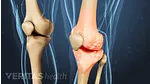
Joint Swelling
People with rheumatoid arthritis usually experience joint symptoms that last 6 or more weeks. Symptoms may include swelling, redness, warmth, pain, and stiffness, particularly after a long period of rest. These symptoms cannot be explained by another condition, such as osteoarthritis or gout.
Blood Tests
While no single blood test can be used to definitively diagnose rheumatoid arthritis, several blood tests can help measure inflammation that may be associated with the disease.
Blood tests used to help diagnose or rule out rheumatoid arthritis include tests that detect:
- Rheumatoid factor (RF)
- Anti-cyclic citrullinated peptide (called ACPA or anti-CCP)
- Inflammatory markers such as erythrocyte sedimentation rate (ESR) and C-reactive protein (CRP)
Blood tests that measure body-wide inflammation can be used to diagnose other diseases, too. For example, CRP may be used to diagnose heart disease.
Imaging Techniques
A doctor may order x-rays, ultrasound, MRI, or other medical imaging to check for signs of RA. Imaging may be especially helpful if the in-office evaluation and lab tests do not provide enough information to diagnose or rule out rheumatoid arthritis.
Ultrasound
This imaging technology is used to check for inflammation of synovial tissues, the delicate membranes that surround many joints and tendons.
- The inflammation of the synovial tissue that surrounds a joint is called synovitis. This inflamed tissue may develop into pannus, a hallmark sign of RA.
- Inflammation of the synovial tissue that surrounds a tendon is a type of synovitis called tenosynovitis. Research suggests tenosynovitis in the finger may be an early sign of RA. 3 Hmamouchi I, Bahiri R, Srifi N, Aktaou S, Abouqal R, Hajjaj-Hassouni N. A comparison of ultrasound and clinical examination in the detection of flexor tenosynovitis in early arthritis. BMC Musculoskeletal Disorders. 2011;12:91. doi:10.1186/1471-2474-12-91.
Ultrasound is performed using either traditional “b-mode” (gray scale) or power Doppler. While less common and more expensive, power Doppler can detect the flow of blood, allowing a physician to see if the synovial inflammation is active.
Ultrasound can also be used to monitor a patient’s response to treatments, though it is rarely used in clinical practice.
X-ray
In the early stages of rheumatoid arthritis, a person may only have joint swelling, which is not detectable on an X-ray, so X-rays are not typically used for early diagnosis.
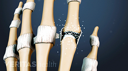
X-rays are typically more helpful after RA has progressed. X-rays can help detect:
- Bone damage (erosions) that occurs as a result of long-standing rheumatoid arthritis
- A narrowing of a joint’s space, which occurs when cartilage degrades and the bones in the joint get closer together
RA bone damage occurs at the joint. For example, if RA affects the knee, the top of the tibia (shin bone) and bottom of the femur (thigh bone) may be damaged. The other parts of the bones are not directly affected by RA.
Eroded bones will look irregularly shaped and/or shaded on an x-ray. If joint cartilage is also lost, the two bones will appear to be closer together than expected—they may even be touching.
Magnetic resonance imaging (MRI)
An MRI is not usually necessary for diagnosing RA. One is typically ordered only if x-rays and ultrasound have not been helpful.
Like ultrasound, MRI can detect inflammation and other changes in the joint’s soft tissue before bone erosion takes place. In addition, an MRI can show bone damage. The drawback to MRI is that it is more time-consuming and expensive than either ultrasound or X-ray.
Physical Exam and Medical History
The appointment will likely begin with the physician taking a thorough medical history. This will include asking the patient several questions, such as:
- What joints are affected?
- How would you describe the pain? (For example, is the pain dull or sharp? Constant or intermittent?)
- Do you have morning stiffness?
- Have you noticed increased fatigue or weight changes?
- Have you experienced other symptoms besides joint pain?
- When did symptoms begin?
- How have symptoms changed over time?
- What movements or activities make the patient feel better or worse?
The history will also include a review of the patient’s:
- Other medical problems
- Previous illnesses and treatments
- Current medications
- Family medical history
See Lifestyle Factors and Fatigue Associated with Rheumatoid Arthritis (RA)
During the physical exam, a doctor will evaluate the patient’s general health and then examine the joints, looking for signs of joint inflammation. For example, a doctor may measure a joint’s range of motion, press the skin over a joint to see if it causes pain, and test joint strength.
- 1 Riaz, S and Kontzias, A. Rheumatoid Arthritis. In: Efthimiou P, ed. Absolute Rheumatology Review. Springer Nature Switzerland AG; 2020; chap 15. Accessed September 15, 2020. https://doi.org/10.1007/978-3-030-23022-7_15
- 2 The European League Against Rheumatism (EULAR). 2010 ACR/EULAR Classification Criteria for Rheumatoid Arthritis (slide 31 of 53). http://www.eular.org/myUploadData/files/RA%20Class%20Slides%20ACR_Web.pdf Accessed August 25, 2016.
- 3 Hmamouchi I, Bahiri R, Srifi N, Aktaou S, Abouqal R, Hajjaj-Hassouni N. A comparison of ultrasound and clinical examination in the detection of flexor tenosynovitis in early arthritis. BMC Musculoskeletal Disorders. 2011;12:91. doi:10.1186/1471-2474-12-91.