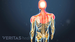
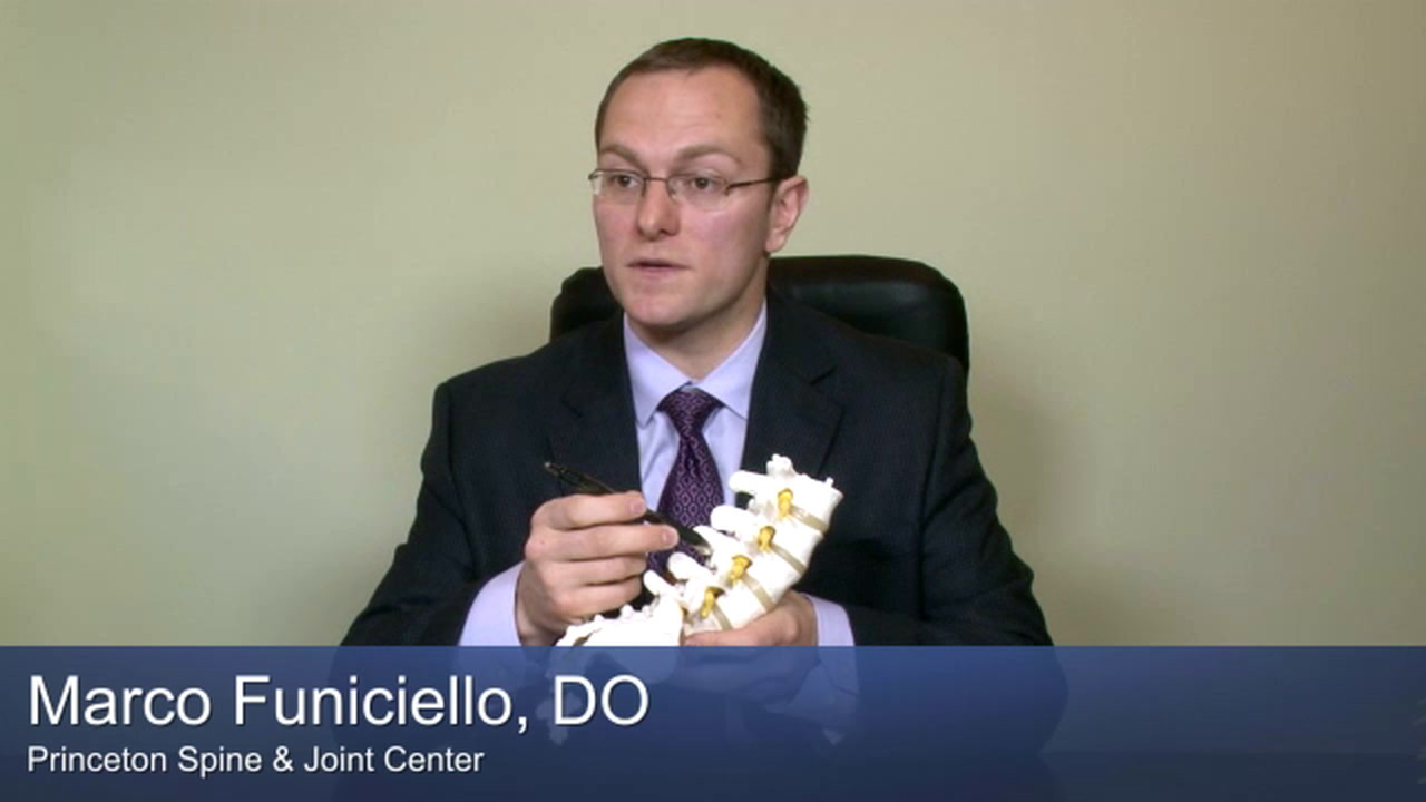
No single test can definitively diagnose osteoarthritis of the spine. A physician must perform a comprehensive evaluation and rule out other causes of back pain, such as a herniated disc, spondyloarthritis, infection, vertebral fracture, and abnormal growths, such as cysts and tumors.
Below is a description of the methods physicians use to make an accurate diagnosis.
In This Article:
Patient Interview
A patient’s reported symptoms are important for diagnosis and treatment. During the interview, a doctor may ask a patient to describe:
- The onset of their symptoms
- The pattern of neck or back pain and stiffness
- How symptoms affect lifestyle
- What eases pain or makes it worse
- Their family medical history
Before the doctor’s appointment, it is often beneficial for a patient to keep a journal to record symptoms and when they occur.
Physical Exam
During the physical exam, a physician will evaluate the patient’s overall health and spine. The exam may include some combination of the following tests:
- Visual inspection of the overall posture and skin overlying the affected area
- Hands-on inspection by palpating for tender areas and muscle spasm
- Range of motion tests to check mobility and alignment of the involved joints
- Segmental examination to check each spinal segment for proper motion
- Neurological examination, including tests of muscle strength, skin sensation, and reflexes
Other maneuvers and special tests may be used to help narrow down the source of pain. Like a patient interview, a physical exam is essential for diagnosing back or neck pain. After these two steps, a physician may be able to give a diagnosis, or they may order additional testing.
Medical Imaging and Lab Testing
Follow-up medical imaging and lab testing may be ordered to provide further information about the extent of the facet joint arthritis and/or to rule out other possible causes of the patient’s pain.
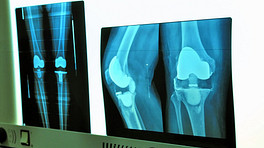
X-rays
An x-ray can show if there is a loss of joint space between the vertebrae, indicating a thinning of vertebral discs. An x-ray can also sometimes show bone spurs at a facet joint, a sign that the bones have tried to compensate for cartilage loss with extra bone growth.
There is a high concentration of nerves radiating from the spine, so even minor cartilage damage or bone spurs detected on an x-ray can translate into a lot of pain if it is in a sensitive spot. It is also possible for a person to experience no pain even though their x-rays to show significant signs of spinal osteoarthritis. Therefore, x-rays are just one tool to be used in conjunction with the patient interview and physical exam.
CT scan
Because of the spine’s complex anatomy, doctors often need a more detailed image of the spine than an X-ray can provide. A computed tomography (CT) scan is essentially many different x-rays layered together to show a cross-sectional view of the spine. For example, CT scans can show whether the spinal canal provides adequate space for the spinal cord.
A CT scan requires a patient to be still for several minutes. A CT scan delivers radiation to the patient—many times more than a single x-ray—so a health care provider may limit the number of CT scans a person undergoes over time.
MRI
Like CT scanning, magnetic resonance imaging (MRI) gives doctors a cross-sectional view of the spine. Unlike an x-ray or CT scan, an MRI does not deliver radiation to the patient, it only uses a strong magnet that provides a detailed look at soft tissues, including vertebral discs, ligaments, tendons, and muscle.
Most MRI scans require the patient to lie flat in a long, narrow tube for 30 to 60 minutes. 1 Medline Plus. Lumbar MRI Scan. US National Library of Medicine. Page last updated August 4, 2020. Accessed August 11, 2020. https://medlineplus.gov/ency/article/007352.htm In some cases, the patient may be standing up.
SPECT scans
A single-photon emission computed tomography (SPECT) scan, uses nuclear medicine and provides visual images of metabolic functions, such as blood flow. It is often used in conjunction with a CT scan to help narrow down specifically where a problem is occurring in the spine.
Read more about Diagnostic Tests on Spine-health.com
Diagnostic injections
A doctor may inject a local anesthetic into the joint or near a nerve that seems to be the source of pain and then observe how it affects the patient’s pain levels. This procedure helps pinpoint the source of pain and can help rule out or confirm certain diagnoses.
- Diagnostic injections are performed by physicians, such as physiatrists, anesthesiologists, neurologists, or neuroradiologists.
- When performing a spinal injection, the physician must use fluoroscopy or ultrasound. These medical imaging tools help the doctor visualize exactly where the diagnostic injection must be placed and ensures that the injectant is not inadvertently placed in a nerve or blood vessel or other unintended structure in the spine.
- Fluoroscopy provides real-time x-ray images. This type of imaging uses contrast dye, which is injected into the patient.
- Ultrasound may be used for certain injections instead of fluoroscopy to eliminate risks related to radiation.
- A diagnostic injection contains a local numbing agent (anesthetic). It may also include a corticosteroid.
- The local numbing agent usually numbs the area for a few hours. If complete pain relief is experienced during this time, then the injected area is the likely source of pain.
- The corticosteroid is designed to have a time-released, anti-inflammatory effect. It may provide some longer-lasting pain relief.
- Because diagnostic injections may help relieve symptoms, they can also be considered therapeutic.
If the person experiences only partial relief from the local anesthetic, then there may be an additional source of pain. Additional injections or diagnostic studies may be considered.
Lab tests
While spinal osteoarthritis cannot be detected through lab tests, they may be used to rule out other conditions, such as spondyloarthritis, infection, or malignancy.
Lab tests may require a blood draw or an aspiration of the spinal fluid (sometimes called a spinal tap). Like diagnostic injections, aspirations of the spine are typically done by a neuroradiologist.
A Comprehensive Diagnostic Approach
To accurately identify the source of back pain, physicians take a comprehensive diagnostic approach. While diagnostic studies (such as an X-ray or MRI) can help verify or rule out a diagnosis, these studies alone, without a patient interview and physical exam, do not provide a conclusive diagnosis.
- 1 Medline Plus. Lumbar MRI Scan. US National Library of Medicine. Page last updated August 4, 2020. Accessed August 11, 2020. https://medlineplus.gov/ency/article/007352.htm